Nanoscopy User Group
 New group name! The STED User Group has become the Nanoscopy User Group
The focus of the Nanoscopy User Group is to create a collaborative space where researchers of all career stages can share their expertise using the latest super-resolution technologies. Sessions will be held on a variety of topics tailored to the needs of the audience, including best practices in experiments and data processing.
Next Meeting : 2026.11.12
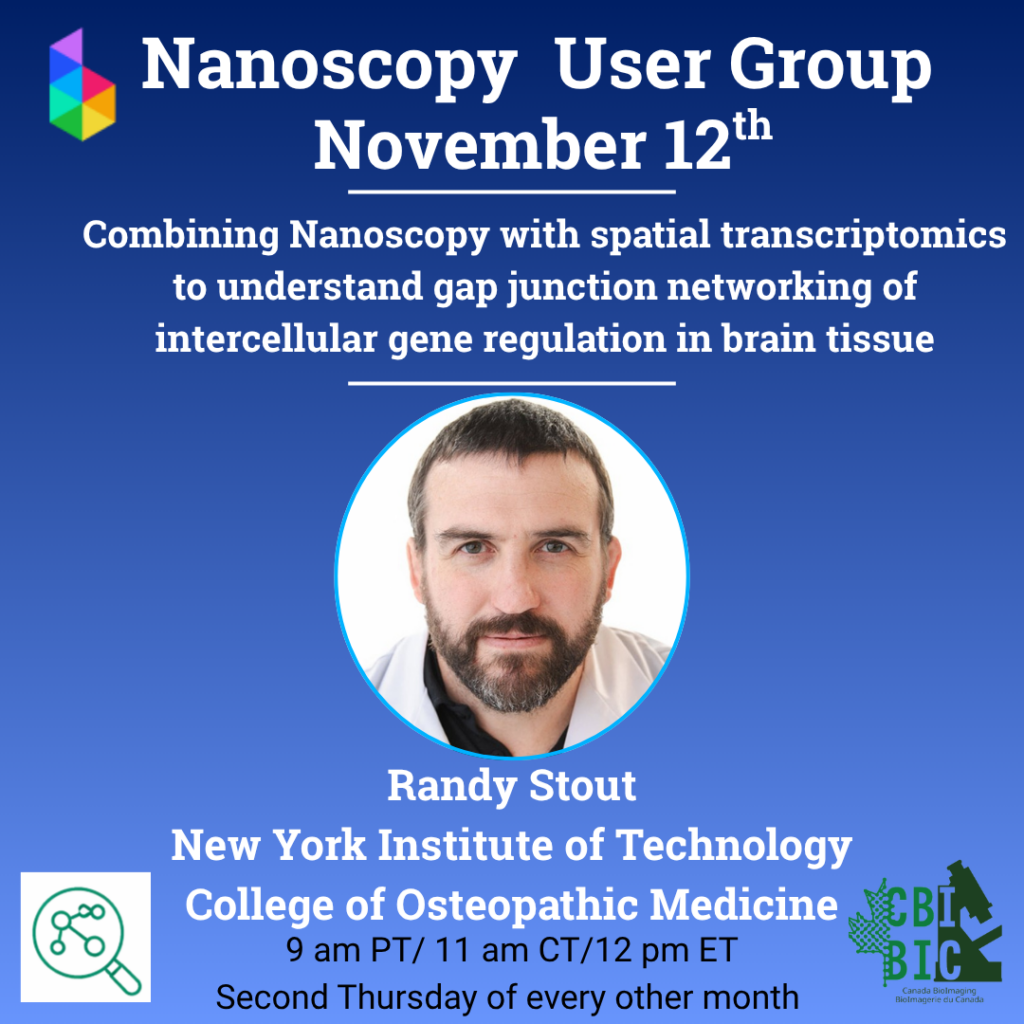
“Combining nanoscopy with spatial transcriptomics to understand gap junction networking of intercellular gene regulation in brain tissue” – Randy Stout, New York Institute of Technology
Talk Abstract: I have been extending the utility of high-plex spatial transcriptomics by adding multi-channel immunostaining and other protein and lipid labeling tools with 3D super-resolution imaging (STED, SMLM, and array confocal). I will present our methodologies that I have developed in this area so far. The specific application for my lab’s research is to reveal the effects of gap junction networking of astrocytes in the brain on intercellular gene regulation.
Randy Stout is an Associate Professor at the New York Institute of Technology College of Osteopathic Medicine, Scientific Director of the Biomedical Research Innovation and Imaging Center (BRIIC), and the Director of the Imaging Center at New York Tech. My lab uses super-resolution microscopy to study nanoscale tissue morphology, gap junctions, astrocytes and thyroid hormone signaling in the brain. I use live-cell, high-resolution laser scanning confocal microscopy and super-resolution microscopy to reveal novel and fundamental aspects of the gap junction plaque structure (quasi-crystalline 2D protein arrays) and anatomy of other supramolecular cellular assemblies. I developed new optogenetic tools and methods for studying fluorescent protein tagged gap junction components during my graduate and postdoc research. Our recent work has provided evidence that gap junction endocytosis can facilitate intercellular mitochondria transfer that complemented parallel work using serial section electron microscopy in other labs.
Questions? bioimagingna.nanoscopyug@gmail.com
User Group Leads


Watch Past Meeting Presentations on YouTube Here!
Past Events
2026.09.10
“Imaging with the MIRAVA POLYSCOPE” – Nicolas H. Piguel, Northwestern University
2026.06.11
“Decoding Nanoscale Protein Organization in NK Cells Using DNA-PAINT” – Hijab Fatima, Columbia University
2026.04.09
“STED workflows for synaptic nanoarchitecture in brain” – Thomas A. Blanpied, University of Maryland
2026.02.12
“MINFLUX Microscopy in a Core Facility: Early Experience and Considerations” presentation by Christina Pyrgaki & “Nanoscopic imaging of structural synaptic changes linked to endogenous hormonal secretion rhythms in the rodent hypothalamus” presentation by Thomas Stroh
YouTube Video link
2025.12.11
Ready, STED, Go: A full-stack workflow from fluorophore to feature extraction” Presentation by Priyam Banerjee
2025.09.11
“A community approach to STED resolution benchmarking using Fourier Ring Correlation (FRC)” Presentation by Henrietta “Henny” Bennett, followed by panel discussion with Joel Ryan & James Jonkman
2025.06.12
“STED, MINFLUX, and their combinations” Presentation by Zachary Connerty-Marin (University of Vienna)
2025.04.10
“Expanding Insights: Magnified Analysis of the Proteome Across Imaging Platforms.” Presentation by Mathias Delhaye & Jeffrey LeDue
2025.02.13
“Investigating Caveolin-1 Endocytosis in Response to Osmotic Shock Using STED Super-Resolution Microscopy” presentation by Timothy Wong
2024.12.12
“Implementing Super Resolution in Core Facilities: Challenges and Opportunities” presentation by Pina Colarusso
2024.10.10
“Multicolor STED Nanoscopy” presentation by Andréanne Deschênes
2024.08.08
Community-Run Discussion
2024.06.13
Kick-off and presentation from Dr. Guang Gao
Partners